| Short Training Programs in Neurosurgery and Otolaryngology |
The Rhoton Neurosurgery and Otolaryngology Surgical Anatomy Program at Mayo Clinic, Rochester, Minnesota offers theoretical and practical training, research fellowships and educational programs in surgical anatomy to improve the teaching-learning process in Neurosurgery, Otolaryngology, and related fields in the US and across the world.
Surgical education, research and surgical innovation are the three fundamental pillars of our mission. As Dr. Mayo stated, “There is no excuse today for the surgeon to learn on the patient”, which makes surgical training an essential part of this program.
Our laboratory follows the well-known and wide-spread foundations and teachings proposed by Albert. L. Rhoton Jr. MD, one of the fathers of modern micro neurosurgery who started his career as neurosurgery staff in Mayo Clinic, Rochester from 1966-1972 before he became Chairman at university of Florida, Gainesville, Fl. Professor Rhoton stated that to perform “accurate, gentle and safe” neurosurgical procedures, neuroanatomy needs to be comprehended. Meticulous dissections and new and safer techniques developed in the surgical anatomy laboratory have a direct applicability in improving patient care in the operating room.
We produce high-quality professional anatomical images of anatomical dissections using 3D-photodocumentantion and post-processing photography techniques (e.g. High Dynamic Range – HDR), which result in peer reviewed publications, books and lectures worldwide.
Microscopic and endoscopic dissections as well as fiber tract dissections are performed in the laboratory. Three-dimensional printing, photogrammetry and virtual reality are increasingly important capabilities of our laboratory.
Microsurgical Anatomy
The understanding of the surgical anatomy of the region to be operated and its surrounding structures is extremely important. This knowledge will aid in choosing the correct surgical approach for a patient and minimize complications. Improvement of approaches, indications, and techniques are one of our goals along with surgical training.
Endoscopic Anatomy
Endoscopic Endonasal Approaches are important procedures in certain regions of the skull base that are accessible through the nose and paranasal sinuses. It is essential for the surgical team have an in-depth knowledge in this region and to develop surgical skills to navigate this complex anatomy.
True translational surgical projects developing new and safer approaches in the laboratory have been of paramount importance to develop this field in the past decade.
Three-dimensional printing and virtual reality
In cooperation with the 3D Anatomy Printing Laboratory directed by Jonathan J Morris, MD the surgical cases and anatomical dissections can be rendered and translated into virtual reality, where the individual anatomy can be studied from any device in a three-dimensional representation or stereoscopic reality. This has improved training and preparation for surgical cases.
Our Team
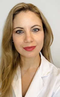
 Maria Peris-Celda MD, PhD – Director
Maria Peris-Celda, MD, PhD is a Professor of Neurosurgery and Associate Professor of Otolaryngology and Anatomy at Mayo Clinic and is a fellowship-trained Skull Base Oncology Neurosurgeon.
She graduated with the highest honors at the University of Valencia, Spain, where she met Professor Martinez-Soriano, her anatomy mentor. She trained in Neurosurgery at la Fe Hospital in Valencia and Albany Medical Center, Albany, NY. She spent two years learning and improving her surgical skills as a fellow of microsurgical and endoscopic neuroanatomy with Professor Albert L Rhoton Jr. MD, from 2011 to 2013 in the Dr. Albert L. Rhoton Surgical Anatomy Laboratory at the University of Florida Gainesville - FL. During these years, she performed most of the dissections and organization of the book “Rhoton’s Atlas of Head, Neck, and Brain. 2D and 3D Images”, which was published by Thieme in 2018 since then has been translated into Korean, Chinese and Portuguese. This book is considered a “must-have” book not only for neurosurgeons and otolaryngologist but also to medical students and any other surgeons who operate and access the head and neck anatomical regions.
“[...] this book is an irreplaceable teaching and professional tool for all spectrum of neurosurgeons and a must-have book in their library [...] all its pictures attest the highest possible quality of specimen preparations, blood vessel injections, impeccable and skillful surgical anatomic dissections, and color photography. [...] particular credit goes to Dr. Maria Peris-Celda and Professor Francisco Martinez-Soriano who worked hard for more than five years to fulfill the promise given to Dr. Rhoton and deliver this work for publication. [...]” Neurosurgery, 83 (6); E232-E233.
Before joining the neurosurgery team at Mayo Clinic, Dr. Peris-Celda worked at Albany Medical Center as a Neurosurgeon where she established the ‘Northeast Surgical Anatomy Laboratory’ where she trained residents and numerous international and national fellows.
Dr. Peris-Celda and her team have been constantly and extensively publishing papers, chapters and books, and is recognized as an international surgical anatomy speaker.
 Luciano CPC Leonel PhD – Laboratory manager and coordinator
Luciano CPC Leonel, received his PhD in Human Anatomy from University of São Paulo – Brazil, and is an Assistant Professor of Neurosurgery. His thesis characterized and described in detail the features an ultrastructure of the venous communication between the cavernous sinus and the pterygoid plexus responsible for venous drainage from the intracranial cavity to the extracranial space. During his PhD he performed a 6 month-fellowship at the Northeast Surgical Anatomy Laboratory – Albany, NY, under Dr. Maria Peris-Celda’s mentorship where he learned and mastered anatomical techniques for preparation of specimens for neurosurgical dissections, microsurgical dissections techniques, 3-D photodocumentation and post-processing and 3D display presentations. One of the papers published by him, Dr. Peris-Celda and her team received the ‘Rhoton Award’ at the 30th North American Skull Base Society Annual Meeting that took place in San Antonio Texas, 2020. He also helped to develop an Educational Program at Albany Medical Center aiming to train and offer a continuing education in the neuroanatomy field for Residents in Neurosurgery collaborating not only with the theoretical lectures but also providing mentorship and guidance inside the lab during the practical tasks proposed by the program.
Luciano Leonel, PhD, has published numerous papers as co-author throughout the past years under supervision of Maria Peris-Celda MD, PhD and Carlos D. Pinheiro-Neto MD, PhD.
| Co-directors | ||
|---|---|---|
 | Matthew L Carlson, MD | Otolaryngology, Otology |
 | Katie van Abel, MD | Otolaryngology, Head and Neck Surgery |
 | Robert J. Spinner, MD | Neurosurgery, Peripheral Nerve |
 | Giuseppe Lanzino, MD | Neurosurgery, Neurovascular |
 | Benjamin D. Elder, MD PhD | Neurosurgery, Spine |
Research fellows
Past fellows
Edoardo Agosti, M.D.
Natalia Rezende, M.D.
Larissa Vilany, M.D.
Danielle Dang, M.D.
Andre Oliveira, Ph.D.
Pedro Plou, M.D.
Giada Garufi, M.D.
Jenna Meyer, M.D.
Simona Serioli, M.D.
Alessandro De. Bonis, M.D
Mariagrazia Nizzola, M.D.
Yasaman Alam, M.D.
Amedeo Piazza, M.D.
Fabio Torregrossa, M.D.
Sheng Chen, M.D.
Current fellows
Megan Bauman, M.S.
A. Yohan Alexander, B.A.
| Programs Offered | |
|---|---|
| CME courses | Mayo Clinic Open and Endoscopic Techniques in Cranial Base Surgery Course July 17 - 20, 2024 |
 | Notify me when registration for the Rhoton Neurosurgery and Otolaryngology Surgical Anatomy Program courses |
Our Research Fellowship Program is offered throughout the year and we accept fellows for a minimum length of stay of 6 months. The lab does not offer personal funding, but we provide the anatomical and technical material needed to complete your training and research projects. The fellowship is mainly directed to neurosurgeons and otolaryngologists but anatomists and other medical professional who perform surgeries in the head, neck and brain areas might be considered. We provide not only structural support but also individual mentorship and guidance during the training for each fellow. Following the methodology of Professor Albert L. Rhoton’s laboratory MD, used in his surgical anatomy lab (Florida-Gainesville), our fellows will have experience with different steps for neuroanatomy investigation from the step-by-step preparation of specimens to post-processing photography techniques and 3D displays. Those interested in applying for our Research Fellowship position should fill out this form, stating your goals and fields of interest, period of time planned to be attended as a fellow in our lab, your filiation, academic titles, updated curriculum vitae and one letter of recommendation. Generally, we take no more than one or two business days to reply our emails. | |
Short Training Programs in Neurosurgery and Otolaryngology - Rochester, Minnesota
| This program is an extended training format dedicated to trainees and neurosurgeons or otolaryngologists who intend to spend from 2-4 weeks developing, improving, and refining their dissection skills and practicing specific and complex neurosurgical or otolaryngology approaches inside our laboratory. The lab offers openings for those planning to attend and perform dissections under guidance of our lab staff. No requirement prior experience regarding dissection, photography techniques or post-processing of images will be necessary. The program will provide specimens and necessary supplies for the dissections. The practice will take place in the laboratory from Monday to Friday during business hours (8am to 5pm).
All the dissections will be mentored by a PhD anatomist or an attending surgeon. |
Coaching and Preparation for the ABNS Neuroanatomy Examination for PGY-2 ( for Mayo and non- Mayo US neurosurgery residents) | |

 Facebook
 X
X LinkedIn
 Forward